
Frequently Asked Questions: LeucoScreen
FAQ1: Will LeucoScreen be available in the future?
No, the product will be discontinued on January 1st, 2020. The kit is replaced by the LeucoScreen Plus kit, which is equally suitable to distinguish between peroxidase-positive and -negative round cells. Moreover, LeucoScreen Plus does not contain a carcinogenic substrate.
FAQ2: In some samples the formation of bubbles makes it almost impossible to read the results under the microscope. What can I do to avoid this ?
The formation of bubbles is part of the chemical reaction taking place between the peroxidase and the hydrogen peroxide. The formation of bubbles is therefore correlated to the amount of peroxidase in the sample. To avoid bubbles from interfering with the interpretation of the results, cover the semen/LeucoScreen mixture as soon as possible after mixing and read the results immediately.
FAQ3: Can I test if the reagent of the LeucoScreen kit are still active ?
Yes you can !
Materials required for the test:
LeucoScreen test
Peroxidase from horseradish (e.g. Sigma code P8125)
Method (the test is performed at room temperature):
- “Activate” 1mL of LeucoScreen dye (Reagent 1) by adding 30uL of hydrogen peroxide (Reagent 2) – to save reagent you may also use 0.33mL of reagent 1 and 10uL of reagent 2 or any other volume as long as the relative amounts stay the same
- Dissolve 1mg of peroxidase into 1mL of demineralized or highly purified water, depending on the concentration indicated on the label of the bottle of peroxidase this will yield a concentration of between 50-150 U/mL of peroxidase
- Add 10uL of the above peroxidase solution to the activated LeucoScreen reagents and mix well
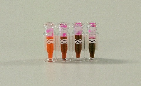
- Depending on the concentration of peroxidase in the initial dilution, the color of the LeucoScreen reagent will change from orange-pink to dark brownish-red within 2-4 minutes. The formation of a precipitate after a few minutes is normal
==> The color change indicates that the reagent is still active
The image below gives an idea of the color change that should be expected from the QC test. The left vial shows the activated LeucoScreen reagent, next are the color changes after 1, 2 and 3 minutes of exposure to the peroxidase.
FAQ4: What is the clinical threshold?
According to the WHO, the threshold is 1×10⁶ peroxidase positive cells/mL. However, this threshold is under debate, as some have found this value too low and others too high.
Threshold levels from 0.2×10⁶ up to 2×10⁶ peroxidase positive cells/mL have been reported. If the threshold is exceeded, additional tests can be done to investigate for accessory gland infection.
FAQ5: How is the concentration of round cells calculated?
Count the number of round cells whilst determining the sperm concentration in a routine semen analysis. If the concentration of round cells exceeds 1×10⁶ cells/mL, additional testing with LeucoScreen is recommended.
FAQ6: Is differentiation between different peroxidase-negative cells possible?
No, this is not possible. Spermatids and spermatocytes can be differentiated with Spermac Stain or a Papanicolaou staining.
FAQ7: How is a pink cell with yellow/brown spots classified?
Classify as “positive”. These cells contain peroxidase that stays inside the granulocytes. If the cells are fully yellow/brown, the granulocytes are no longer intact.
