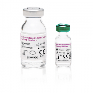
Hyaluronidase in FertiCult Flushing medium
80IU hyaluronidase solution in FertiCult Flushing medium
Hyaluronidase in FertiCult Flushing medium contains 80 IU/ml hyaluronidase from bovine origin, in FertiCult Flushing medium (HEPES-buffered medium which contains 4.00g/liter human serum albumin). The product is ready for use. Information about the composition of the product can be found in the material safety data sheet.
Hyaluronidase in FertiCult Flushing medium is used in the oocyte denudation process. Hyaluronidase digests the hyaluronic acid between the cumulus cells, which makes it easier to remove the cumulus mechanically.
Regulatory
Europe: CE-marked – Brazil: registered
Product order codes
HYA001 : Hyaluronidase solution in Flushing Medium – 5x 1 mL
HYA010 : Hyaluronidase solution in Flushing Medium – 10 mL
pH | 7.3-7.6 |
Osmolality | 270-290 mOsm/kg |
Sterility | Sterile |
Endotoxin | < 1.0 EU/mL |
Mouse embryo assay | ≥ 80% blastocysts after 96h incubation (30 minute exposure at 1-cell stage) |
Albumin | FDA (USA) and EMA (Europe) compliant |
Shelf life | 12 months from date of produce |
Component | Benefit |
HEPES (18 mM) | HEPES stabilizes the pH under air (Clark and Swain 2014), therefore CO2 incubation is not required. |
HSA (4 g/L) |
Hyaluronidase in FertiCult Flushing medium contains 4 g/Liter Human Serum Albumin (HSA) to optimize medium performances. HSA is universally added to most ART media because it is widely considered to be of benefit.
The role of albumin in ART media is extensive, including: |
Hyaluronidase (80IU/ml) | Hyaluronidase in FertiCult Flushing medium contains bovine derived hyaluronidase (80IU/ml). Hyaluronidase is an enzyme derived from bovine testes and is capable of hydrolyzing hyaluronic acid (Tranchepain, et al. 2006), the latter being one of the most abundant constituents of the extracellular matrix (Laurent and Fraser 1992).
When collected, each oocyte is surrounded by a matrix of cells called the cumulus-oocyte complex (COC). The COC is composed of cumulus granulosa cells embedded in a matrix of long hyaluronan oligosaccharide chains cross-linked by a complex of hyaluronan binding cell surface and extracellular matrix proteins and proteoglycans (Richards 2005). In vivo, enzymes like hyaluronidase are released by the acrosome reaction of the sperm cells and dissolve the intracellular matrix between the cumulus cells. Also in vitro during ART, COC around oocytes are removed (i.e. “oocyte denudation”). For that purpose, the European Society of Human Reproduction and Embryology (ESHRE) recommends to use an enzymatic procedure with hyaluronidase, which facilitates the subsequent mechanical denudation process using a pipette (Magli et al. 2008). |
Aghaways, IHA., KM. Falah and AA. Ali , The difference in the outcomes between surgically retrieved and ejaculated spermatozoa for Intracytoplasmic Sperm Injection Cycles in Sulaimanyah province., Acta Medica International (2016),Vol.3,No.1,pp.30-38
Ciepiela, P., T. Baczkowski, A. Drozd, et al., Arachidonic and linoleic acid derivatives impact oocyte ICSI fertilization-a prospective analysis of follicular fluid and a matched oocyte in a ‘one follicle-one retrieved oocyte-one resulting embryo’ investigational setting., PLoS One (2015),Vol.10,No.3
Desquiret-Dumas, V., A. Clement, V. Seegers, et al., The mitochondrial DNA content of cumulus granulosa cells is linked to embryo quality., Hum Reprod (2017),Vol.32,No.3,pp.607-614
Lédée, N.; Lombroso, R.; Lombardelli, L.; et al., Cytokines and chemokines in follicular fluids and potential of the corresponding embryo: the role of granulocyte colony-stimulating factor., Human Reproduction (2008),Vol.23,No.9,pp.2001-2009
Lédée, N.; Munaut, C.; Sérazin, V.; et al., Performance evaluation of microbead and ELISA assays for follicular G-CSF: a non-invasive biomarker of oocyte developmental competence for embryo implantation., Journal of Reproductive Immunology (2010),Vol.86,pp.126-132
May-Panloup P., Chrétien M.F., Jacques C., et al., Low oocyte mitochondrial DNA content in ovarian insufficiency, Human Reproduction (2005),Vol.20,No.3,pp.593-597
Mitchell V., Steger K., Marchetti C., et al., Cellular expression of protamine 1 and 2 transcripts in testicular spermatids from azoospermic men submitted to TESE – ICSI, Molecular Human Reproduction (2005),Vol.11,No.5,pp.373-379
Qassem, E., K. Falah, I. Aghaways and T. Salih , A Correlative Study of Oocytes Morphology with Fertilization, Cleavage, Embryo Quality and Implantation Rates after Intra Cytoplasmic Sperm Injection., Acta Medica International (2015),Vol.2,No.1,pp.7-13
Sigala, J., C. Sifer, D. Dewailly, et al., Is polycystic ovarian morphology related to a poor oocyte quality after controlled ovarian hyperstimulation for intracytoplasmic sperm injection? Results from a prospective, comparative study., Fertil Steril (2015),Vol.103,No.1,pp.112-118
Vidberg, F.; Zeggari, R.; Pieralli, C.; et al., Measurement of oocyte temporal maturation process by means of a simple optical micro-system., Sensors and Actuators B: Chemical (2011),Vol.157,pp.19-25
Alvarez, JG., and BT. Storey, Differential incorporation of fatty acids into and peroxidative loss of fatty acids from phospholipids of human spermatozoa., Mol Reprod Dev. (1995),Vol.42,No.3,pp.334-46
Blake, D., P. Svalander, M. Jin, et al., Protein Supplementation of Human IVF Culture Media., Journal of Assisted Reproduction and Genetics (2002),Vol.19,No.3,pp.137-143
Clark, N.A. and Swain, J.E., Buffering systems in IVF., Culture media, Solutions, and Systems in Human ART, by P. Quinn (2014),pp.30-46
Laurent, T C., and J R E. Fraser, Hyaluronan, The Journal of the Federation of American Societies for Experimental Biology (1992),pp.2397-2404
Magli MC, Van den Abbeel E, Lundin K, et al., Revised guidelines for good practice in IVF laboratories, Human Reproduction (2008),pp.1253-1262
Malda, J. et al., Cell Nutrition, In Tissue engineering, by C. Van Blitterswijk. London, UK: Academia Press, Elsevier, (2008),pp.327-362
Richards, JS, Ovulation: new factors that prepare the oocyte for fertilization, Molecular and Cellular Endocrinology (2005),Vol.29,pp.75-79
Tranchepain, F., et al., A complete set of hyaluronan fragments obtained from hydrolysis catalysed by hyaluronidase: appllication of studies of hyaluronan mass distribution by simple HPLC devices., Analytical Biochemistry (2006),pp.232-242
Hyaluronidase in FertiCult Flushing medium is a ready-to-use cell culture medium for oocyte denudation